|
types of injuries associated with it. Confusion is often found when using the
term “knee” since a horse has four legs. The stifle is the true “knee” of a
horse and is found only on the hind limbs of the horse. The forelimb of the
horse is comparable to the human arm so the “knee” of these limbs is
structurally similar to our wrist. More properly it should be called the carpas.
 Two
individual joints make up the stifle and it is determined by the bones that join
together there. The femoral-tibal joint is used to communicated between the
large bone in the upper leg called the femur and the smaller bone below it
called the tibia. Communication from the patella or kneecap is sent to the femur
through the femoral-patella joint. The stifle is made up of these two joints. A
thin capsule surrounds the entire stifle joint that has a specialized fluid to
help with shock absorption and lubrication. Two
individual joints make up the stifle and it is determined by the bones that join
together there. The femoral-tibal joint is used to communicated between the
large bone in the upper leg called the femur and the smaller bone below it
called the tibia. Communication from the patella or kneecap is sent to the femur
through the femoral-patella joint. The stifle is made up of these two joints. A
thin capsule surrounds the entire stifle joint that has a specialized fluid to
help with shock absorption and lubrication.
Structural stability is also provided through ligaments in this joint. The
inside and outside of the stifle has specific ligaments that keep the leg from
bending excessively in either direction. These ligaments are referred to as
collaterals and can be either torn or damaged if a horse slips or falls.
There are two large crossing ligaments in the center of the stifle joint. They
form an X inside the joint by attaching to the femur and tibia. These ligaments
also prevent the leg from bending excessively and are called cruciate ligaments.
Cruciate ligaments are easily damaged as anything who has played football, skied
or played any other contact sport knows. The most common sports related injury
today among humans is the tearing of the front or anterior ligament in the knee.
This structure is the same in the horse with the same potential for injury.
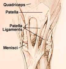
In the front of the thigh the largest muscle is the quadriceps. In humans this
muscle is attached to the kneecap by one thick ligament and another thick
ligament attaches the kneecap to the lower leg bone or tibia.
The same job is done in horses by three patella ligaments that help to make the
horses stifle stronger. While standing this allows the horse to lock their leg
by shifting their weight and rotating the patella so that one of these ligaments
locks over a ridge located on the femur. This is what allows the horse to sleep
while standing with a minimum of energy.
However, this system is always correct in the way it works. Some horses may have
stifles that once locked can’t be suddenly released if they either have very
straight, upright back legs when born or have poor quadriceps muscles.
If a horse shows a slight hitch in their gait then this condition is subtle and
is especially noticed when going downhill. However, the condition can also be
severe when the leg is completely locked out behind the horse with the leg being
unbendable.
This condition is called upward fixation of the patella. There are a number of
treatment options including exercise that includes working up and down gentle
slopes or lunging in sand to more serious treatment options such as injections
along the patella and surgery.
Some additional means of distributing the forces placed on the stifle is
necessary since it is a large joint that carries a lot of weight. Between the
ends of the femur and the tibia there is two thick pieces of C-shaped fibro
cartilage that help act as additional shock absorbers. These helps stabilize the
joint and are known as menisci. The menisci play an important role by reducing
the amount of wear and tear on the cartilage surface of the joint. Falls or
other trauma can tear the menisci which can result in damage and lame horses.
Inflammation and swelling of the stifle joint is likely to result from any
damage to these structures. In jumpers and event horses the most commonly seen
condition is fractures of the patella which can occur after hitting an obstacle
while jumping or from kicks or falls. Fractures on the femur and tibia bones are
less common in horses.
Since the joint contains the majority of the stifle structure it is difficult to
see, touch and evaluate. For veterinarians this presents a diagnostic issue.
Because of the severity of lameness it can usually be easy to diagnose tearing
of the main support ligaments, but for mild sprains and bruising of the
ligaments it can be difficult for a veterinarian to diagnose.
Recently veterinarians are starting to use ultrasound evaluation more often and
it is helping to provide a wealth of new information. However, the horses risk
is increased since there is general anesthesia required and it is also not
regularly used for diagnostic purposes since it is expensive.
Less invasive procedures such as CAT scans and MRIs may become available for
horses in the future and this may allow veterinarians to get a better look at
the stifle joint and unlock some of the mysteries regarding the lameness of this
joint.

|